
101 - 110 of 127
manual oct fundus camera
Selling leads
|
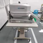
Examination Of Damage To The Optic Nerve Of The Eye SD OCT Machine Compared with the color fundus images of plane imaging, one fundus SD OCT Machine ...
2026-09-18 00:05:18
|
|
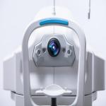
...fundus camera CE DICOM 3.0 Gamma,contrast,brightness,color of image can be adjusted Automated Fundus Camera Five automatic functions 1. Three...
2026-09-18 00:05:18
|
|
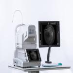
...Fundus Camera non-mydriatic Automated fundus camera RetiCam 3100 can be used to capture the anterior and posterior eye images. It has a field of ...
2026-09-18 00:05:18
|
|
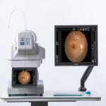
...fundus camera dual camera system intellegent operation Automated Fundus Camera Five automatic functions 1. Three-dimensional automatic tracking. 2. ...
2026-09-18 00:05:18
|
|
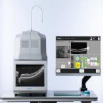
oct scan machine PC inside easy installation white for various fundus diseases and glaucoma oct scan machine is an automatic posterior segment OCT ...
2026-09-18 00:05:18
|
|
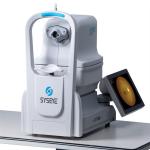
digital eye fundus camera fundus imaging intelligent operation digital eye fundus camera design-minimum pupil size 2.8mm Achieve peripheral retinal ...
2026-09-18 00:05:18
|
|
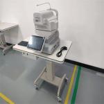
...Fundus Retina Was Expanded Digital Fundus Camera Fundus screening is very important for patients with diabetes and hypertension. In order to ...
2026-09-18 00:05:18
|
|
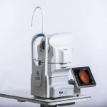
...a field of view of 50 degrees for fundus imaging. If features: Auto focus (AF) and auto shooting (AS) Small pupil imaging capability In default ...
2026-09-18 00:05:18
|
|
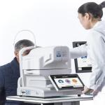
...camera one click automatic FFA small pupil dual camera Automated Fundus Camera Five automatic functions 1. Three-dimensional automatic tracking. 2. ...
2026-09-18 00:05:18
|
|
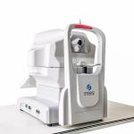
Patient History Data Can Be Browsed Non Mydriatic Fundus Camera Non Mydriatic Fundus Camera design-minimum pupil size 2.8mm Achieve peripheral retinal ...
2026-09-18 00:05:18
|