
21 - 30 of 102
automated non mydriatic fundus camera
Selling leads
|
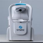
...images of fundus exposed to visible light within milliseconds (no enough time for pupils to shrink). Kestrel 3100m can photography the retina, i.e. ...
2026-07-08 00:18:14
|

|
...Non Mydriatic Fundus Camera is its versatility in fixation options. Users have the choice between external fixation or internal fixation, allowing ...
2025-11-04 21:57:47
|

|
...Non Mydriatic Fundus Camera is its versatility in fixation options. Users have the choice between external fixation or internal fixation, allowing ...
2026-03-15 00:28:15
|
|
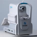
eye fundus camera pupil size of 2.8mm dual camera system DICOM and AI As a key step of diagnosis and treatment, fundus examination plays an extremely ...
2026-07-08 00:18:14
|
|
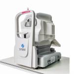
ophthalmic fundus camera Field angle is 50° 220V, 50 Hz. Class I ophthalmic fundus camera is to take photos of the fundus of the eye through a fundus ...
2026-07-08 00:18:14
|
|
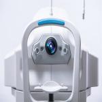
...fundus Auto fluorescence (FAF optional) Automated Fundus Camera Automated Fundus Camera RETICAM 3100 Fundus auto fluorescence (FAF), as a new ...
2026-07-08 00:18:14
|

|
Product Description: Equipped with both auto and manual exposure modes, this fundus camera offers flexibility and control for capturing the perfect ...
2026-04-08 19:07:56
|

|
Product Description: One of the key features of this fundus camera is its flexibility in fixation options. Users can choose between external fixation ...
2026-04-08 19:07:56
|
|
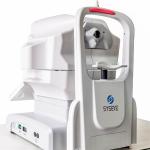
digital eye fundus camera manual/auto DICOM and AI system soft lighting Kestrel 3100m can photography the retina, i.e. neurosensory tissues of eyes, ...
2026-07-08 00:18:14
|
|
Used To Diagnose Any Underlying Pathology Digital Fundus Camera The eye is the only organ in the human body that can observe the microvessels without ...
2026-07-08 00:18:14
|