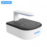
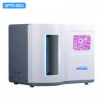
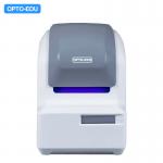
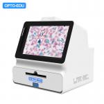
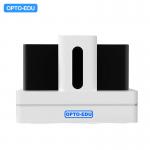
- 5 Standard pathology slides (75*25mm), or 2 Standard large
pathology slices (75*50mm)
- High Quality Plan Apochromatic Objective Lens APO20x N.A. 0.8
- 5.0M Scientific Research-grade Large-area CMOS Digital Camera
- Scanning Resolution 20x Scan ≤0.24um/pixel, 40x Scan ≤0.12um/pixel
- Scanning Speed 15x15mm Area 20x Scan <35s, 40x Scan <50s
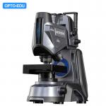
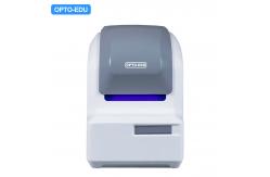
| M30.5815 Full Auto Microscope Slide Scanner Specification | | Slide Capacity | 5 Standard pathology slides (75*25mm), or | | 2 Standard large pathology slides (75*50mm) | | Objective | APO 20x N.A.0.8 high quality plan apochromatic objective lens | | Camera | 5.0M Scientific research-grade large-area CMOS digital camera | | Scanning Method | High-speed continuous area scanning | | Scanning Resolution | 20x scan ≤ 0.24um/pixel | | 40x scan ≤ 0.12um/pixel | | Scanning Speed | 15x15mm scanning area: | | -20x scan, <35 seconds | | -40x scan, <50 seconds | | Scanning Mode | Fast, Accurate, 3D, Fusion mode | | Automatic Identification | Automatic identification of tissue area, label code, character and
slide type | | Real-time Browsing | Observe slide information without scanning, switch zoom ratios,
capture, edit and export high-definition images | | Software | Scan Software: iScanneX, Browser Software: iViewer | | Ai Application | Supports work with Artificial Intelligence software such as
cytology, histology, and immunohistochemistry |
|
