
Autofocus Non Mydriatic Fundus Camera System For Cataracts Check
|
|
advantage of fundus imaging technology with light going through pupils Non Mydriatic Fundus Camera
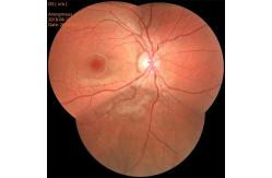
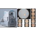
Non Mydriatic Fundus Camera is to examine the vitreous and retina, choroid, and optic nerve disease important check method of fundus camera check is necessary, you can better check out the concrete causes, found the cause, symptomatic treatment can, if you have cataracts in the elderly is treated, if severe cases will affect the normal vision.
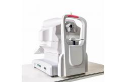
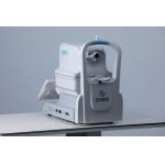
Working principles of Non Mydriatic Fundus Camera RetiCam 3100, based on near-infrared light imaging technology, can capture the required visible light images of fundus exposed to visible light within milliseconds (no enough time for pupils to shrink). RetiCam 3100 can photography the retina, i.e. neurosensory tissues of eyes, and then convert the captured optical image into electronic pulses that can be understood by the brain. Pupils serve as both the inlet and outlet of light for illumination and imaging of RetiCam 3100, thus monitoring the fundus. Patients shall put the chin on the chin rest and lean the forehead on the forehead rest. The operator shall operate it manually to focalize and align to the fundus, and capture the fundus images upon clicking, with such images directly shown on the touch screen. Ophthalmologists will trace the progression of eye diseases on the basis of such retinographs, which will serve as the basis of diagnosis and treatment.
Specification of Non Mydriatic Fundus Camera
Focusing on the two major public health problems of adolescent myopia and elderly ophthalmology, our company researches new diagnostic and treatment equipment for ophthalmology, develops low-cost applicable technology products, realizes industrialization, reduces the cost of social medical and health system, and serves the strategic needs of national health.The company always adhere to the "high-tech,new vision" for the enterprise development concept.We have a strong technical force, has been awarded 11 patents, and obtained 13,485 quality system certification.Bio has established an efficient marketing team and a perfect after-sales service system to provide medical equipment with the highest cost performance and meticulous service. |
||||||||||||||||||||||||||||
| Product Tags: Autofocus Non Mydriatic Fundus Camera Automated Non Mydriatic Fundus Camera FDA fundus imaging system |

|
Non Mydriatic Fundus Camera with 13mm Working Distance ±25D Diopter Compensation and 9-position Mosaic for Retinal Imaging |
|
High-Performance Non Mydriatic Fundus Camera with Dual Camera System for Comprehensive Eye Health Monitoring |
|
Non Mydriatic Fundus Camera With Dual Camera System DICOM And AI For Eye Fundus Diagnosis |
|
Non Mydriatic Fundus Camera With Auto Focus Alignment And Eye Fundus Alignment |
|
Professional Non Mydriatic Fundus Camera For Accurate Imaging |
|
Automatic Retinal Imaging Fundus Camera 135° Wide Field 17mm Working Distance |