
Electromagnetic Ultrasound Diagnostic Machine CE Retinal Detachment Detection
|
|
B Probe Uses Electromagnetic Drive Has A Long Service Life Ophthalmic Ultrasonic Scanrne
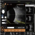
Retinal detachment, choroidal detachment and pigment membrane leakage syndrome can all be detected by Ophthalmic Ultrasonic Scanrne , and can be used to identify the location, extent, shape, and level of detachment. Vitreous hemocele and vitreous machinery can also be found by ultrasonic examination, especially when accompanied by poor lens or vitreous transmittance, the effect of ultrasound can not be replaced by other optical examination methods. Ultrasound examination can determine whether there is a foreign body in the eyeball, can determine the location of the foreign body, the distance from the eye wall, the size and shape of the foreign body (which helps to determine the treatment method), whether the foreign body can move, whether there is combined with retinal detachment, vitreous opacity, and can determine whether the foreign body is magnetic material under ultrasonic monitoring. introduced of Ultrasonic ScannerRetiWave1000 ophthalmic AB ultrasound diagnostic instrument is a complete ultrasound system, which has the following basic functions: 1. A super function is the measurement of eye axis length. 2. B ultrasonic function is fan scan. 3. IOL calculation function USES 6 formulas to calculate the artificial crystal.
Feature:
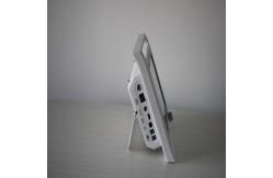
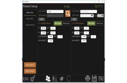
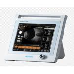
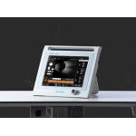
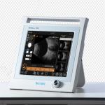
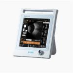
1) 12.1" color and touch screen . 2) Ultrasonic Scanner With the same software as Quantel , top one in China With advanced intelligent digital software and parameters of freezed and Stored images could be adjusted voluntarily. 3) Can have different results on the report according to different constants A Scan A measures data for every part of the eyeball as anterior chamber depth, lens thickness axial length and so on which are needed in ophthalmic surgeries , and calculates IOL by axial length. 4) B scanner use electromagnetic ,better save B probe life . Scan B displays profile images of the eyeball clearly and directly. Scanning anatomical forms and nidi inside the eyeball, doctors can diagnose accurately for examination of cataract , vitreous body disease, ocular trauma,detachment of retina or choroid , macula disease , and intraocular tumor, etc 5) B scanner ,video playbackof 100 images. 6) A scanner , one group with 10 data to get average ,At the same time the accuracy is 0.05mm
Technical Data
SOFTWARE OF Ultrasonic Scanner
PACKAGE
Company Introduction Focusing on the two major public health problems of adolescent myopia and elderly ophthalmology, our company researches new diagnostic and treatment equipment for ophthalmology, develops low-cost applicable technology products, realizes industrialization, reduces the cost of social medical and health system, and serves the strategic needs of national health.The company always adhere to the "high-tech,new vision" for the enterprise development concept.We have a strong technical force, has been awarded 11 patents, and obtained 13,485 quality system certification.Bio has established an efficient marketing team and a perfect after-sales service system to provide medical equipment with the highest cost performance and meticulous service. |
||||||||||||||||||||||||||||||||||||||||||||||||
| Product Tags: 10MHz Ultrasound Diagnostic Machine Electromagnetic Ultrasound Diagnostic Machine CE eye ultrasound machine | ||||||||||||||||||||||||||||||||||||||||||||||||
|
RetiWave1000P Digital Ophthalmic Ultrasound Scanner with Convenient Measurement Tool for Accurate Diagnosis |
|
Automatic Save 4d Ultrasound Scanner Machine 10MHz NonFocusing Imported Probe |
|
Mini Ultrasound Scanner Machine With Intelligent Digital Software Multiple IOL Calculation Formulas |
|
IOL Calculation Ultrasonic Scanning Machine 10MHz Convenient Measurement Tool |
|
110dB Ophthalmic Ultrasonic Scanner Automatic Calculation Of Average |

|
Automatic Save 4d Ultrasound Scanner Machine 10MHz NonFocusing Imported Probe |