
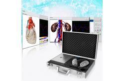
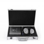
Clinical Metatron NLS Biofeedback Health Analyzer High Precision With OEM
|
|
2019 Biofeedback Health Analyzer NLS 4025 Metatron Hunter Health Detection Machines
Description and Field of Application Telemetric nonlinear analysis data processing apparatus "Metatron" (hereinafter "apparatus") can beused for nonlinear analysis of biological structures and testing of biologically active supplements. The apparatus can also be used in research centers and scientific research facilities.
Main feature for Metatron 4025 Hunter The main feature of Metapathia GR Hunter software is function called “Three-dimensional Scanning” which allows to localize automatically a nidus where tumor appeared,heritable diseases, etc. and to find the reason of appearance at genome level, passing one after another histological cuts, cytological cuts, chromosome sets, separate chromosomes, and go deeper to fragments of DNA helix. nter NL
Etalon testing Every organ and every cell has its own specific and distinctive oscillation. The oscillations are stored in the computer memory and can be displayed on the screen as a graph, which represents the condition of the information exchange between an organ or tissue and the environment. Every pathology has its own individual and distinctive graph. The computer memory also stores a very large number of pathological processes taking into account rate of evidence, age, sex and other variations.
After frequency characteristics are read from tissue, the apparatus compares spectral similarity with stored processes (healthy tissue, pathological tissue, infection agents) and then selects the closest pathological process or tendencies to its appearance. By this method of analysis it is possible to trace the condition of the red (S) input signal and the condition of the blue (N) output signal, which are displayed as graphs on the screen. From the shape of the graph it is possible to determine which of the reference
BIOCHEMICAL HOMEOSTASIS This program carries out a qualitative evaluation of the main biochemical factors by evaluating the wave functions of body tissues. This evaluation is carried out using the NLS – analysis mode. Note that the lowest values of enzyme (hormone) concentration within the normal bounds correspond to 2 in the graph. Whereas, the highest values within the normal bounds correspond to 6.
The values of the factors equal to 3, 4 or 5 correspond to the ‘mode’ of the factor, and the extreme values 1 and 7 characterize biochemical factors beyond the physiological norms, lower and higher respectively. The standard rules for making biochemical analysis using conventional clinical methods should be used when analyzing the computer results.
PATOMORPHOLOGY This shows a list of the etalons of destructive processes. This section holds basic patomorphologic conditions peculiar to single tissues of an organism. Every destructive process has a distinctive graph.
MICROORGANISMS AND HELMINTHS In this section are recorded the major characteristics of infection agents: – bacteria, viruses, mycoplasma, rickettsias, fungi and helminths. This presents changes in the form of high peaks of dissociation within the frequency range representing the natural frequencies of the tissue. For example Opisthorhis felineus has a high dissociation in
frequency – 4.9Hz – parenchymatous liver tissue and bileexcreting
tissue. These tissues are known to be largely affected by
trematodes Opisthorhis felineus-
NUTRICEUTICALS
LITHOTHERAPY
Natural frequencies of the tissues within the following standard frequency band:
– 3.4 unstriated muscular tissue. – 4.2 tessellated epithelium of the digestive tract;
– 4.9 – 5.8 kidney tissue epithelium and reproductive organs;
– 7.4 central sections of sensory analysers except the optic ones,
and sub cortical structures of the brain, pons cerebelli;
cerebellum, limbic system and lungs parenchyma;
By pressing “Display” button following drop list appears: “Object”
– graph lines of lilac and orange color, display a graph of the
examined biological object, organ or tissue plotted in the course
of the investigation.
WHERE IS THE NLS TECHNOLOGY BORN? Nonlinear systems are inspired by the discoveries and ideas of Theodore Van Hoven, who discovers the theory of the logic of quantum entropy, summarizing scientifically “The exchange of information between systems is carried out at a distance in an associative and selective manner, non-linear thanks to the appropriate amount of electromagnetic radiation, which has the adequate energy to destabilize the links of the elemental structure of the system. All this is based on the development of Russian technology and the need to perform high-level non-invasive treatments in the human body.
WHAT IS THE TECHNOLOGY OF BIORRESONANCE OF NON-LINEAR SYSTEMS OR NLS? We will say that it is one of the most up-to-date scientific
advances in the diagnosis and treatment of diseases, and is based
on the scientific evidence that every organism and tissue has its
own electromagnetic frequency, and it is possible to evaluate it
through the exchange of information between non-linear systems. The
intensity of the exchange of information means that it is possible
to establish a connection and frequency exchange with any living
tissue through its electromagnetic frequency.
You can also work with:
WHAT ARE THE MOST ELEMENTAL FUNCTIONS OF THE 4025 HUNTER
WHAT BENEFITS WOULD YOU BUY A HUNTER 4025 18D
Purpose
The Metapathia-GR Hunter software can operate only with the telemetric nonlinear analysis data processing apparatus “Metatron” and its subsequent modifications. The telemetric nonlinear analysis data processing device is compatible with the IBM-type PCs and intended for studying reaction of a biological object to different types of the informational impact. “Metatron” allows correlating the measurement process with the process affecting it and performs the following.
6) It transmits the W values from the unit into PC memory upon completion of the measurements and saves them in unit memory of prior to the beginning of recording data of the next measurements.
The apparatus is intended to register psychophysics changes in system and allows to:
Special requirements
Operating principle and operational procedures
The sensor detects faint signal fluctuations and selects them from the average statistical noise characteristics of the field and converts them to a digital sequence that is processed by a microprocessor, which is then transmitted to the computer through the interface cable.
Capacity and productivity
The system is designed to diagnose one patient at a time. The operating cycle takes from 3 minutes to 1 hour. The system can run non-stop for 24 hours. The computer, according to the established program operates automatically in adjusting and controlling all information. The results of the patient’s diagnosis are displayed on the monitor screen and kept on a separate file on the hard disk. The information can then be transferred to an individual diskette for future use. The current information is displayed on the screen as required.
Also you can:
-Green line shows there is no pronounced functional change in
evidence. Press ‘Exit‘ to exit the program.
Some of the many functions performed with this machine.
EXPLAINING SOME EXAMPLES:
Researches In the Ultra-structure mode histological, cytological and molecular researches are carried out. Moving the cursor over organ’s projection, you select the structure of your interest, which image and name appear on the right, and the cursor becomes a cross-hair. Clicking the left mouse button starts researches of a respective structure at the preset point. After the researches are completed an icon emerges on the organ’s image, which can be unfolded by clicking it with the left mouse button. On one and the same organ you can make several researches of ultra-structures with different localizations, gradually passing from microscopic sections to cytological preparation and molecular structures.
Types of researches:
The form “Researches” allows to carry out computer nonlinear analysis in the mode of a programmed and (or) individual selection of the organs intended for researches. The main feature of “Metapatia-GR Hunter” software is “3D scan” function, which allows automatically localize the nidus where tumor and hereditary diseases appeared, find out the reason of appearance on genome level, passing one by one histological, cytological preparations, chromosomes and going deeper to the level of DNA molecule parts.
You can turn on this mode by pressing “3D scan” button. If this button is not pressed, researches will be carried out in normal mode, without localizing of nidus where pathology appeared. During the research the most grave changes in tissues shown on macrocuts are revealed, then search and research of histological cuts of these tissues in areas of the most significant pathological changes are carried out. Then, after histological cut research, the search for the most significantly changed cells is carried out, which examined to reveal changes in cell structures. After that, the algorithm of the search goes to the level of chromosomes, to estimate the changes in single chromosomes, then it goes deeper to the level of DNA molecule research. During the research in “3D scan” mode the estimation of topological picture and metastasis is carried out. Additional researches fulfilled to search metastases into other organs. The research is done in automatic mode; user can observe it and stop at any moment.
The button is multi-purpose for starting of the research, estimation of micro- and additionally placed points, nidus estimation and making of preparation. The name of the button is changed according to fulfilled function.
The button “Analysis” allows carrying out a routine analysis based
on the results of the researches.
The switch-button “Programmed/individual choice“; when it is pressed allows an automatic selection of a profound detailed researches of anatomical, histological and cytological structures depending upon the presence of frank changes in complete anatomical sections of the body; when the button is not pressed the physician can solely select for researches the organs belonging to one of the anatomical systems, by putting or removing tags on the organ’s picture, in the right part of the screen on the split bar with right mouse button.
The button “Make preparation” – the preparation is made automatically for pictures, in which the nidi were evaluated. After clicking this button the operator should select the organs subject to preparation making and press the button “Start preparation making”.
In organs catalogue there are two modes of representation: -graphical – organs shown as pictures; -textual – shown names of organs. You can switch modes by pressing “Textual mode” button. In textual mode “Sort” and “Cancel/Restore chosen” buttons are available. “Sort” button allows change modes of organs list sorting. There are four modes: -According to systems. Organs sorted according to systems: main catalogue, digestive system, respiratory system, urogenital system, cardiovascular system, blood and lymph, endocrine system, nervous system, sensors and musculoskeletal system.
-According to points. In the beginning of the line shown organs with more pronounced changes in estimation points. The ”Options button” shows additional buttons panel.
CLOSING SOME CONCEPTS OF ANALYSIS The buttons under the organ’s picture can be either depressed or pushed up; in the depressed condition organ’s respective elements are depicted. “Text” – allows acquiring information on particular fragments in the picture. To this effect you have to press the “Text” key, which will display icons in the picture, shaped as green daggers. To read the text, stop the mouse cursor on the dagger, and then a message will appear in the square next to it. Left-clicking the dagger se nds the message to print.
To this effect put ticks in squares to the left from the text of the message, in the unfolded form. In this mode there is a possibility to quick-cross for examining the picture connected with this particular research with the help of icons.
To this effect press the “Icons” key, this will display icons in the picture. In order to see what a picture a particular icon can unfold, you need to stop the mouse cursor on the icon, then the picture name will appear in a square next to it, and in place of the graph there will be its image with icons. The color square around the icon denotes the functional condition of the organ.
The color of the square corresponds to the colors described in “Program setup” section; clicking an icon unfolds the respective organ for researches
CONTACT US! More professional introduction for your better reference ! Send your inquiry details in the below, click ‘Send’ now ! We will reply you and slove your consideration within 24 hours !
|
||||||||||
| Product Tags: metatron nls diagnostic body health machine |

|
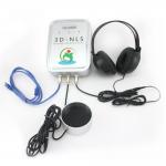
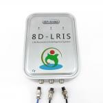
Windows OS Biochemical Analysis 8D LRIS NLS Diagnostic Device |
|
Silver Color Metatron NLS Full Body Health Analyzer High Accuracy CE Certification |
|
Biochemical Analysis Body Health Machine 95% Accuracy Nls Health Analyzer |
|
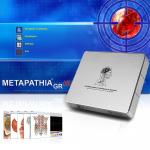
Medical Health Diagnostic Device Biofeedback Metapathia Nls 4025 Hunter |
|
Clinical Examination Aids Full Body Health Analyzer 8d Nls Health Analyzer |
|
8D NLS Health Analyzer Machine Stable Chakra Aura Machine Body Detection |