| Specification | A63.7140 | A63.7160 | | Key Parameters | Resolution | 1.4nm@15kV(SE) 2.5nm@30.0kV(BSE) | 0.9nm@30kV(SE)
1.2nm@15kV(SE)
1.5nm@1kV(SE, BD mode) | | Accelerating Voltage | 0.02kV~30kV | 0.02kV~30kV | | Magnification | 1~2000000x | 1~2000000x | | Electron Gun | Schottky Thermal Field Emission Gun | Schottky Thermal Field Emission Gun | | Probe Current | 1pA~40nA | 1pA~40nA | | Field of View | 6mm | 6mm | | Dwell Time | 20ns | 20ns | | Beam Deflection | -- | Dual Beam Deflection System:
Electromagnetic & Static Hybrid Beam Deflection System | | Objective Lens | Dual Objective System:
Magnetic Objective Lens & Electrostatic Objective Lens,
Magnetic Sample Adaptable | Dual Objective System:
Magnetic Objective Lens & Electrostatic Objective Lens,
Magnetic Sample Adaptable | | Gun Aperture | (10μm,30μm,70μm,100μm,150μm,220μm)*2 Sets (1 For Backup), Motorized
Moving | (10μm,30μm,70μm,100μm,150μm,220μm)*2 Sets (1 For Backup), Motorized
Moving | | Chamber | Chamber Size | Width 370mm, Height 330mm, Depth 344mm | Width 370mm, Height 330mm, Depth 344mm | | Extension Port | 10 Ports | 10 Ports | | Vaccum System | 2 Ion Pump
1 Turbo Molecular Pump
1 Mechanical Pump Oil Free | 2 Ion Pump
1 Turbo Molecular Pump
1 Mechanical Pump Oil Free | Gun Vacuum: 2x10⁻⁷Pa
Chamber Vacuum: 6x10⁻⁴Pa | Gun Vacuum: 2x10⁻⁷Pa
Chamber Vacuum: 6x10⁻⁴Pa | | Stage | 5 Axes Auto Stage, X:130mm, Y:130mm, Z:60mm, R: 360°, T: -10°~70°, Maximum load >500g | 5 Axes Auto Stage, X:130mm, Y:130mm, Z:60mm, R: 360°, T: -10°~70°, Maximum load >500g | | Camera | Optical Color Navigation CCD
High Definition IR CCD | Optical Color Navigation CCD
High Definition IR CCD | | Detectors & Extensions | Standard | SE Detector | SE Detector
Inlens SE Detector | | PC & Software | Computer | Working Station, Memory 16G, Hard Disk 512G, 24" Monitor, Win10
System | Working Station, Memory 16G, Hard Disk 512G, 24" Monitor, Win10
System | | Control | Control Panel & Joystick | Control Panel & Joystick | | Software | Auto Focus, Auto Stigmator, Auto Brightness Contrast, Image Format
TIFF,JPG,PNG,BMP, Image Output Resolution Max 16k*16k | Auto Focus, Auto Stigmator, Auto Brightness Contrast, Image Format
TIFF,JPG,PNG,BMP, Image Output Resolution Max 16k*16k | | Optional Accessories | A50.7101 | BSE | BSE | | A50.7102 | - | InLens BSE | | A50.7103 | Energy Dispersive Spectroscopy (EDS/EDX) | Energy Dispersive Spectroscopy (EDS/EDX) | | A50.7104 | Electron Backscatter Diffraction Pattern (EBSD) | Electron Backscatter Diffraction Pattern (EBSD) | | A50.7105 | EDS+EBSD | EDS+EBSD | | A50.7106 | Scanning Transmission Electron (STEM) | Scanning Transmission Electron (STEM) | | A50.7107 | Electron-beam-induced Current (EBIC) | Electron-beam-induced Current (EBIC) | | A50.7108 | Cathodoluminescence (CL) | Cathodoluminescence (CL) | | A50.7109 | Plasma | Plasma | | A50.7110 | Air Lock, Sample Exchange Warehouse | Air Lock, Sample Exchange Warehouse | | A50.7111 | Beam Blanker | Beam Blanker | | A50.7120 | Large lmage Stitching Software | Large lmage Stitching Software | | A50.7121 | Particle Analysis Software | Particle Analysis Software | | A50.7112 | Vacuum Transfer Holder | Vacuum Transfer Holder | | A50.7113 | Raman-SEM Correlative System | Raman-SEM Correlative System | | A50.7115 | UPS | UPS | | A50.7114 | - | Column built-in energy fiter ExB |
▶ Strong Compatibility, High Adaptability Can be installed on different terminals, such as computers, mobile
phones, and tablets, to control the electron microscope; This
SEM-OS electron microscope operating system is compatible with SEM
from various manufacturers and is compatible with multiple models,
expanding the SEM ecosystem ▶ Integrated Software and Computing, Simple and Efficient Unified user interface, no need to repeatedly adapt to different
terminals; Equipped with AI algorithms to collect information and
present real-time output effects with clearer image quality and
more prominent details; Kernel driven SEM accelerates hardware
control |
① Menu bar, ② Quick operation area, ③ Data bar, ④ Monitoring area,
⑤ Navigation area, ⑥ Comprehensive area, ⑦ Operation area, ⑧ Status
area |
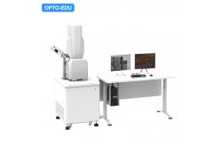
The A63.7140/A63.7160 series scanning electron microscope is
equipped with IGS vacuum transfer rods, EDS energy spectrometers,
Raman spectroscopy and other accessories, providing a comprehensive
solution for lithium battery research from sample preparation,
morphology observation, composition analysis, and structural
analysis. |
Step 1:The transfer rod is loaded onto the glove box to complete the
sample transfer from the glove box to the transfer rod compartment. Step 2:The sample transfer process involves transferring the positive
pressure inside the rod chamber during the transfer process. Step 3:The transfer rod is loaded onto the electron microscope to transfer
the sample from the transfer rod chamber to the main chamber of the
electron microscope. Step 4:Sample shooting and data post-processing, customized development
according to user needs. ▶ SEM+ EDS Spectrometer + Vacuum Transfer Rod + Raman Spectroscopy +
Analysis Software Structural analysis | Mechanism analysis | High precision
displacement table ▪ Make up for the molecular structure analysis that EDS
technology cannot achieve, and comprehensively grasp the sample
composition ▪ Realize fast switching between Raman optical axis and
electron beam optical axis, multi-dimensional analysis of sample
characteristics, and real-time tracking. The structural evolution of materials during charging and
discharging processes, and the in-depth study of their assisting
mechanisms ▪ Large stroke high-precision high-speed piezoelectric
ceramic displacement table, achieving integrated data acquisition
at the same position, meeting Stability analysis of long-term
confocal Raman surface |
|