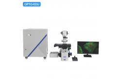
Laser Confocal Microscope, Full Auto Motorized- Laser 405nm, 488nm, 561nm, 640nm, Detector AOTF Wavelength
400-750nm, 4 PMT
- Scan Mode X-Y,X-Y-T,X-Y-Z, X-Y-Z-T, Hexagon Shape Pinhole,
Continuouslv Variable Transmission(CVT)
- Confocal Field Number Square Inscribed in Dia.18mm Circle, Image
Bit Depth 16bits
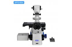
- A16.1098 Full Motorized Inverted Fluorescent Microscope X/Y/Z
Working Stage 325x144mm Moving Range 130x100mm
- Full Set Including Computer + 4K Monitor + Digital Camera,
Professional 3D Software
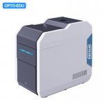
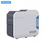
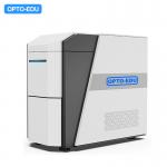
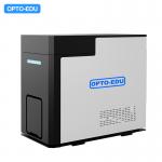
Laser Confocal Microscope A64.1095
It Is Used For Accurate Imaging Of Biological Slices, Living Cells
Or Internal Structures Of Living Tissues; Three-dimensional Image
Reconstruction Analysis; Multi-channel Fluorescence Channel
Analysis, Fine Analysis Of Spectral Signals; The Qualitative,
Quantitative And Localization Distribution Of Biological Substances
Such As Molecules, Organelles Or Ions Are Detected. |
| Optical Path Design of A64.1095 The laser output of all lasers is controlled by the acousto-optic
controller (AOTF). After integration, it enters the scanning head
system and can be turned on with one key to avoid the risk of
cross-color caused by multiple channels and ensure the stability
and accuracy of the optical path output. |
Interactive Operation Convenient interactive mode and multiple control methods could meet
different needs of users from beginners to professional users.
Combined with the powerful features for software and hardware
interactive automation of this product , it has greatly simplified
the whole Set experimental process , which could easily realize
generation of three-dimensional structure and analysis functions
such as time-lapse Analysis of multiple regions etc. By using
matched NOMIS Advanced C. |
High signal-noise ratio High resolution image
Obtaining high signal-noise ratio images based on high-sensitivity
photo multiplier light(PMT) and stable laser light source
At the same time , the system adopts high-speed scanning
galvanometer to realize real-time scanning up to 4096x4096
Resolution, the use of large numerical aperture
objective(100times,N.A=1.45) ensures high-quality imaging
resolution. |
| A64.1095 Laser Confocal Microscope, Full Auto Motorized | | Confocal Laser Unit | | Laser Unit | 4 Laser Units:
Laser 405 nm Optical Fiber Export Power 30mW, End Power 16mW
Laser 488 nm Optical Fiber Export Power 30mW, End Power 16mW
Laser 561 nm Optical Fiber Export Power 30mW, End Power 16mW
Laser 640 nm Optical Fiber Export Power 30mW, End Power 16mW | | AOTF | The Laser Ooutput of All Lasers Is Controlled By the Acousto-Optic
Tunable Filter (AOTF). After Integration, Lasers Enter The Scanning
Head System And Can Be Turned On With One Key, Avoiding The Risk of
Cross-Color Caused By Multiple Channels And Ensuring The Stability
And Accuracy of The Optical Path Output.
Laser Intensity Adjustment Range 0.01%-100%,
Minimum Adjustment Step Accuracy 0.01% | | Detector | Wavelength 400-750nm, High Sensitive 4 PMT, One of PMTs Is Used For
640nm Channel And DIC Channel Switching | | DIC Detector | Wavelength 400-750nm, High Sensitive 1 PMT | | Scanner | The Confocal Scan Head Is Coupled to The Left Interface of The
Microscope Body to Achieve The Highest Quality Optical Path
Imaging.
Maximum Pixel Size: 4096x4096, 4K Real Time
Scanning Speed:
2 FPS(512 x 512) ,
8 FPS(256 x 256) ,
0.5 FPS(1024×1024) ,
0.12 FPS(2048×2048),
0.03 FPS(4096×4096) | | Scan Mode | X-T, Y-T, X-Y, X-Y-Z, X-Y-Z-T | | Pinhole | Hexagon shape, Continuouslv Variable Transmission (CVT), Adjust
Range 0~0.5mm | | Field Number | Confocal Scan Field: Square Inscribed In Dia.18mm Circle (14x14mm) | | Image Bit Depth | 16 Bits | | Motorized Inverted Fluorescent Microscope (A16.1098) | | Optical System | NIS60 Infinite Optical System (F200) | | Eyepiece | EW10x/25mm, EP17.5mm, Adjustable Diopter -5~+5°, Dia.30mm | | Head | Seidentopf Trinocular Head, Inclined at 45°, Interpupillary
Distance 47-78mm, Eyepiece Tube Dia.30mm,Fixed Visibility; Light
Split Switch E100/P0,E50/P50,E0/P100, Built-in Bertrand Lens
Position Adjustable | | Output Port | Splitting Ratio: Left:Eyepiece=100:0; Right:Eyepiece=100:0 | | Nosepiece | Motorized Sextuple Nosepiece, With DIC Slot, M25x0.75 | | Objective | NIS60 Infinity Plan LWD APO Objective, Cover Glass 0.17
APO 10x N.A.0.45, W.D. 4.0mm
APO 20x N.A.0.75, W.D. 1.1mm
Semi-APO 40X N.A.0.95, W.D. 0.3mm
APO 60x N.A.1.42, W.D. 0.14mm,Oil
APO100x N.A.1.45, W.D. 0.13mm, Oil | | Condenser | 6-Position Motorized Condenser, N.A.0.55, W.D.26, Slot For Phase
Contrast Plate 10x/20x, 40x, 60x Optional, Slot For DIC Plate 10x,
20x/40x Optional | | Illumination | Transmitted Kohler Illumination10W LED | | Epi-Illumination Wide-Field Fiber Illumination, With 6-Position
Motorized Fluorescent Disc, Including B,G,U Fluorescent Filters,
With Motorized Fluorescent Shutter | | Intermediate | Manual 1x, 1.5x, Confocal Switching | | Workig Stage | X/Y/Z Motorized Working Stage 325x144mm, Moving Range 130x100mm,
Maximum Speed 25mm/s, Resolution 0.1μm, Repeat Accuracy 3μm, With
Mechnical Adjustable Slide Clamp | | Focusing | Manual & Motorized Coaxial Coarse and Fine Focusing Adjustment,
Focusing Stroke Up 7mm, Down 2mm, Coarse Stroke 2mm/Rotation, Fine
Stroke 0.002mm/Rotation, Minimun Stroke 0.01um Under Motorized
Control | | DIC | DIC Plate 10x, 20x, 40x Plate, Can Be Inserted in Nosepiece Slot,
Optional | | Controller | Joy Stick Controller, Control Box, USB Cable | | Computer + 4K Monitor + Digital Camera | | Computer | 1. Windows 10 Pro 64 bit Operating System | | 2. CPU: Intel Core i7-8700, 6 Core, 12MB Cache, 3.20GHz, 4.6Ghz
Turbo w/ HD Graphics 630 | | 3. RAM: 16GB (2x8GB) 2666MHz DDR4 UDIMM Non-ECC | | 4. Hardware: 3.5"" 1TB 7200rpm SATA Hard Disk Drive | | 5. Video card: NVIDIA Quadro P620, 2GB, 4 mDP to DP Adapter | | 6. USB Interface: 6 Available USB Slots | | 7. Display: 24” Monitor Display that Supports 1920X1080 Resolution | | Software | NOMIS Advanced Version, Display/Image Processing/Analysis 2D/3D/4D
Analysis, Time-lapse Aanalysis, 3D Volume Render/Orthogonal, Image
Stitching, Multi-channel Color Confocal Image | | Camera | USB 3.0 Digital Camera For Fluorescent Image |
|